Table of Contents
Washington University Experience | NEOPLASMS (NEURONAL) | Papillary Glioneuronal Tumor (PGNT) | 2A4 Papillary Glioneuronal Tumor (PGNT, Case 2) smear 3.jpg
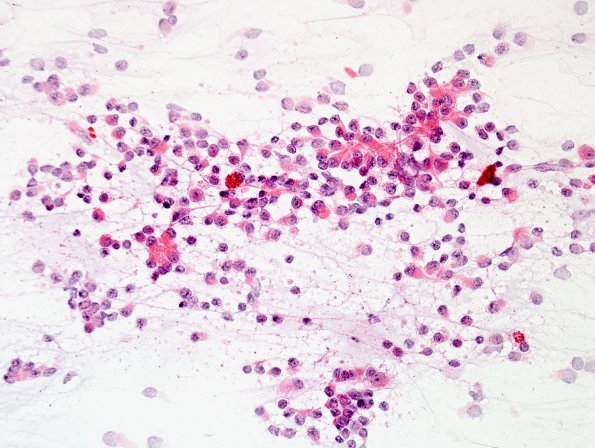
Other portions of the intraoperative smear show a neoplasm composed of cells with bland, round to oval nuclei and a moderate amount of eccentrically situated eosinophilic cytoplasm. In many of the papilla, there are detectable eosinophilic elongated processes extending from the surrounding cells towards the central blood vessel. (H&E)

