Table of Contents
Washington University Experience | NEOPLASMS (NEURONAL) | Papillary Glioneuronal Tumor (PGNT) | 2H1 Papillary Glioneuronal Tumor (PGNT, Case 2) EM 1 copy - Copy
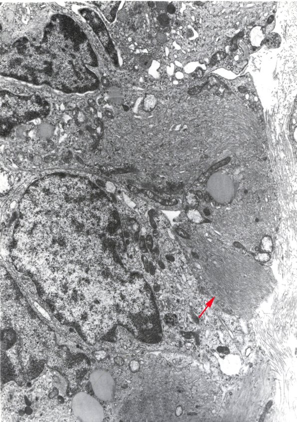
Ultrastructural examination reveals numerous cells containing large bundles of intermediate filaments as well as occasional cellular inclusions resembling dense core granules. Not seen are definite microvilli or cilia, intracellular microlumina, or zipper-like junctions typical of an ependymal neoplasm. Synaptic junctions were not found. (electron micrograph)

