Table of Contents
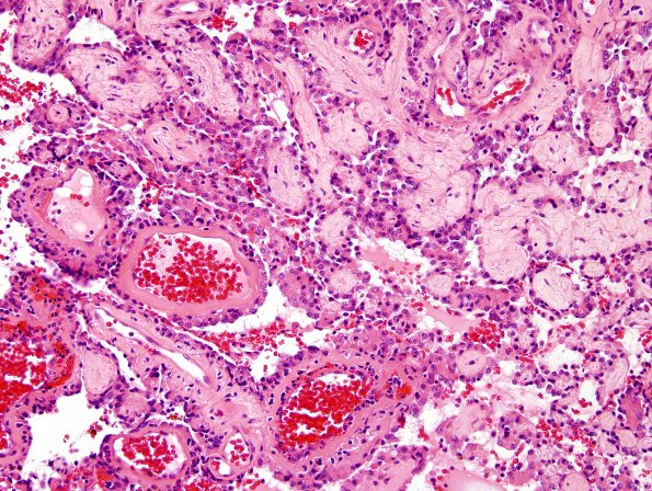
Washington University Experience | NEOPLASMS (NEURONAL) | Papillary Glioneuronal Tumor (PGNT) | 3A9 Papillary Glioneuronal Tumor (PGNT, Case 3) H&E 17.jpg
In some areas, cuboidal tumor cells are seen lining hyalinized vessels with free cells between papillae. Mitotic figures are hard to find and there is no evidence of endothelial hyperplasia or necrosis. (H&E)

