Table of Contents
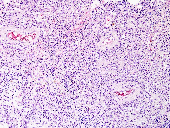
Washington University Experience | NEOPLASMS (NEURONAL) | Papillary Glioneuronal Tumor (PGNT) | 5A3 Papillary Glioneuronal Tumor (PGNT, Case 5) H&E 4.jpg
Sections of the right parietal biopsy show a proliferation of pseudopapillary structures, composed of hyalinized blood vessels, surrounded by perpendicularly oriented cells with delicate fibrillary processes; between the fibrovascular cores, cells with a similar appearance to those seen surrounding blood vessels are growing in a sheet-like pattern. (H&E)

