Table of Contents
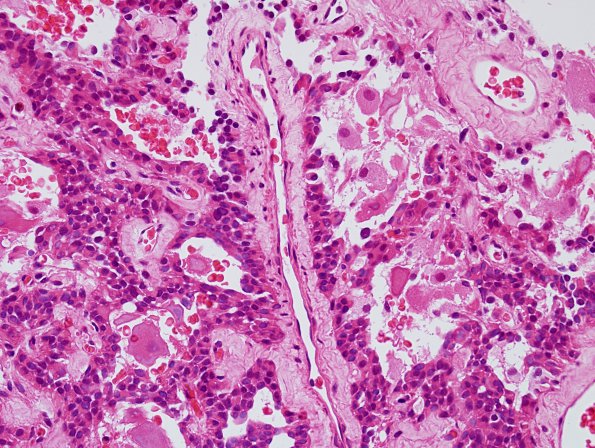
Washington University Experience | NEOPLASMS (NEURONAL) | Paraganglioma - Cauda Equina Neuroendocrine Tumor | 11A1 Paraganglioma GGL Diffn (AANP 2003 Case 2) H&E 1.jpg
Case 11 History (AANP 2003-Slide 2) ---- The patient was a 47-year-old female who developed menorrhagia and abdominal, pelvic, and back pain in February 2002, with associated 50-pound weight loss. Pelvic examination revealed an enlarged uterus. Ultrasound reportedly showed a heterogeneous echogenic mass in the uterine body. MRI was performed on 3/14/02 and showed a heterogeneous mass in the anterior myometrium that measured 10.5 x 10.5 x 9.5 cm and was felt to be suspicious for an atypical leiomyoma or sarcoma. Also present was an intradural, extramedullary enhancing mass at the level of L3. The patient was examined by a neurosurgeon, who elicited a history of intractable back and bilateral leg pain, but neurological examination was normal. Operation on the spinal mass revealed a 4 x 3 x 2 cm reddish-gray encapsulated tumor within the center of the neural canal at the level of L3. The caudal pole of the tumor was noted to have a fine band of tissue exiting from it that was felt to be the filum terminale. Multiple venous vessels and a single arterial vessel were noted to enter the dorsal and rostral aspect of the tumor. Gross total resection was achieved without loss of lumbosacral nerve roots.

