Table of Contents
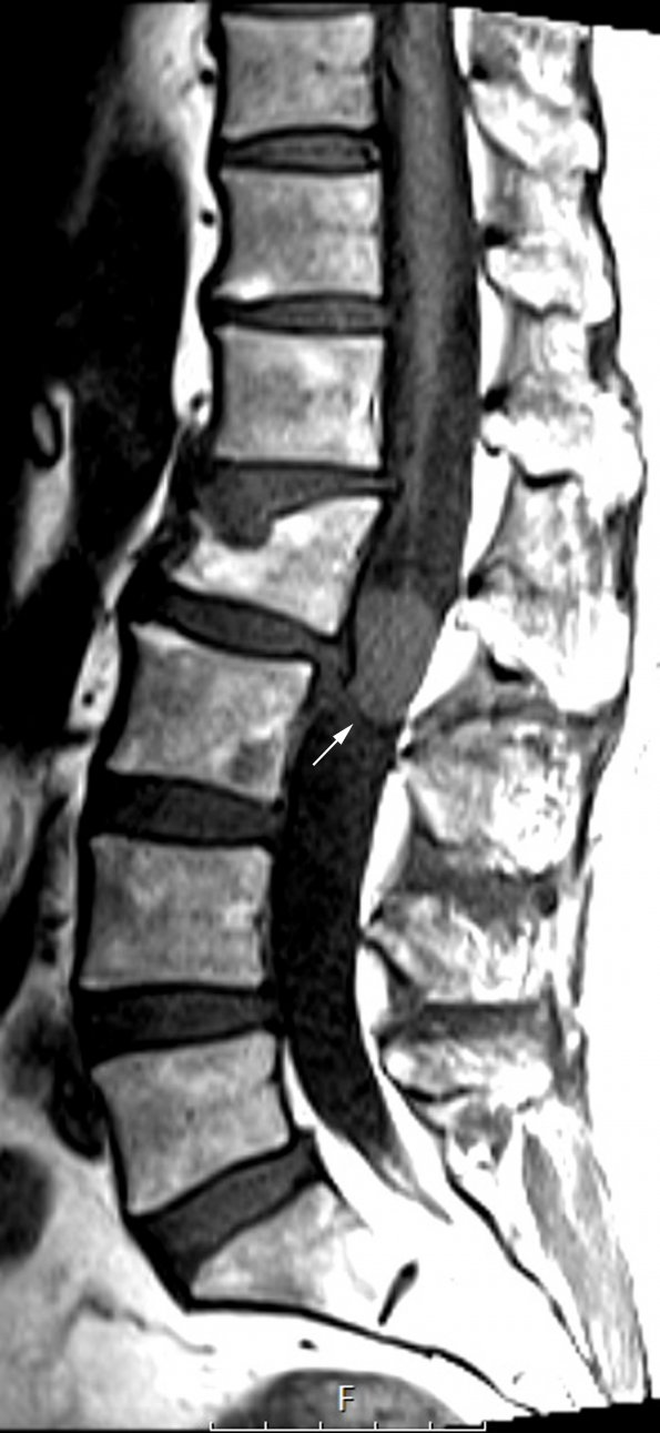
Washington University Experience | NEOPLASMS (NEURONAL) | Paraganglioma - Cauda Equina Neuroendocrine Tumor | 1A1 (Case 1) T1 No C copy - Copy
Case 1 History ---- The patient was a 59 year old woman with several weeks of radiating right leg pain. A spine MRI at an outside institution showed a 2.0 x 1.5 x 2.0 cm intradural, extramedullary, avidly-enhancing mass at L2. Operative procedure: L2-L3 laminectomy with intradural tumor resection. ---- 1A1-3 Spinal MRI studies: 1A1,2 A discrete mass (arrow, 1A1) is shown in the L2-3 space in this T1-weighted element without (1A1) and with contrast administration (1A2) resulting in tumor enhancement.

