Table of Contents
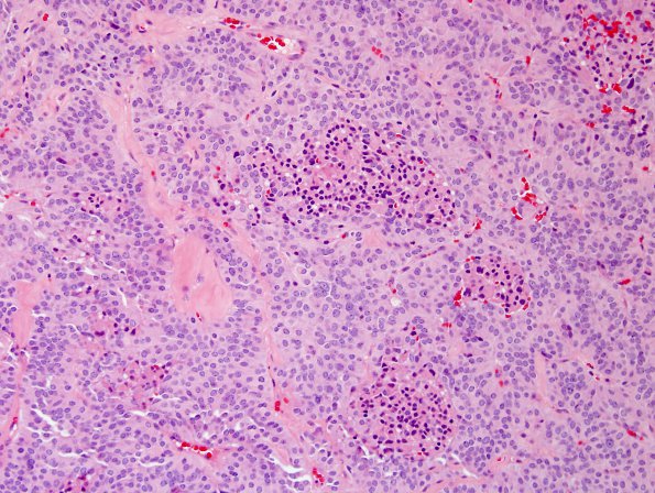
Washington University Experience | NEOPLASMS (NEURONAL) | Paraganglioma - Cauda Equina Neuroendocrine Tumor | 9A1 Paraganglioma (Case 9) H&E 2.jpg
Case 9 History ---- The patient was a 72 year old female who underwent back surgery for spinal stenosis in 2003. Over the last 6 months in 2006, she developed sensory symptoms in the legs, and imaging revealed a solid mass at the L5 level. ---- 9A1-3 Microscopic sections reveal a paraganglioma. Tumor cell density is moderate, and a neuroendocrine pattern is seen with tumor cells arranged in cords, ribbons, and irregular clusters; the classic 'zellballen' pattern is only focal at best. In 9A1 there are scattered areas of necrosis and pyknotic nuclei. Perivascular pseudorosette-like structures are seen focally and vascularity is dense. Several blood vessels are hyalinized. Ganglion cell differentiation is not seen. Tumor nuclei display little atypia and are round to oval and contain small nucleoli; associated eosinophilic cytoplasm is moderate in amount. Mitoses are uncommon.

