Table of Contents
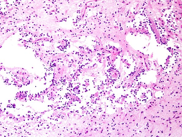
Washington University Experience | NEOPLASMS (NEURONAL) | Rosette-Forming Glioneuronal Tumor (RGNT) | 1B1 RGNT (S05-35206) H&E.jpg
1B1-4 H&E sections from this brain biopsy material show moderately cellular tumor tissue, with a fine fibrillar parenchyma interrupted by mucin-filled microcystic spaces. Oligodendroglia like small round nuclei, with fine chromatin and perinuclear clearings decorate the borders of microcysts and fine vessels. Relatively dense finely fibrillar eosinophilic columns are distinctive, yielding many neurocytic rosettes when sectioned perpendicular to the longitudinal axis. (H&E)

