Table of Contents
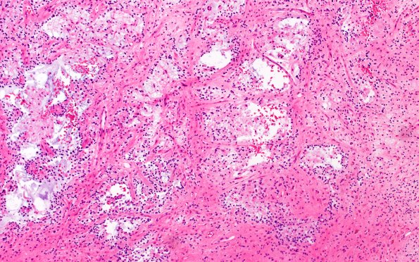
Washington University Experience | NEOPLASMS (NEURONAL) | Rosette-Forming Glioneuronal Tumor (RGNT) | 5A1 RGNT (Case 5) H&E 8
Case 5 History ---- The patient is a 45yo male with a history of hypertension and ruptured splenic cyst; he then presented with 10 days of diplopia. Brain MRI showed a cystic non- or minimally-enhancing left inferior cerebellar mass. Operative procedure: Posterior fossa craniotomy for left cerebellar tumor resection. ---- 5A1 Routine H&E stained sections show a low-grade neoplasm with predominance of glial elements resembling pilocytic astrocytoma. The glial component is composed of mostly compact fibrillary areas with "piloid" (hair-like) processes and some loose areas with mucinous stroma harboring cells with an "oligodendroglial" phenotype. Abundant areas of dystrophic calcifications and hemosiderin deposits are identified admixed within the tumor. Numerous blood vessels in the background show marked hyalinization of their walls. Microvascular proliferation and necrosis are not appreciated. Fragments of adjacent cerebellar folia show partial to near complete replacement by tumor.

