Table of Contents
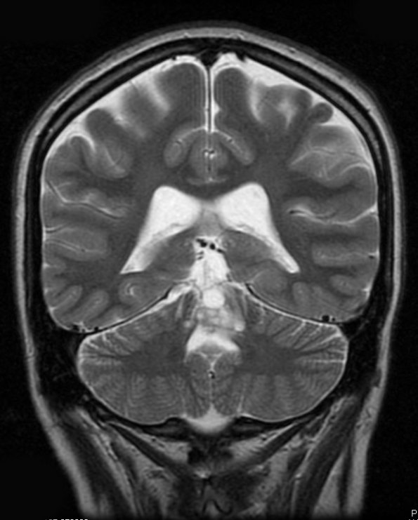
Washington University Experience | NEOPLASMS (NEURONAL) | Rosette-Forming Glioneuronal Tumor (RGNT) | 6B2 RGNT (Case 6) T2 - Copy
The scans are all T2-weighted showing MR imaging performed at BJH in January 2015 shows postoperative changes in the posterior fossa as well as a focus of enhancement at the superior vermis measuring 1.0 x 1.2 cm. The focus of enhancement is associated with increased cerebral blood volume. Additionally, the vermis appears cystic with multiple punctate foci of enhancement. There is increased FLAIR signal at the superior vermis and tectum. Operative procedure: Midline suboccipital posterior craniotomy for resection of tumor with use of Stealth frameless sterotaxy, microsurgical dissection, and IMRI.

