Table of Contents
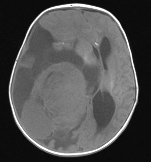
Washington University Experience | NEOPLASMS (NON-GLIAL NON-NEURONAL) | Choroid Plexus Carcinoma (CPC) | 1A1 Choroid Plexus Carcinoma (Case 1) T1 1 - Copy
Case 1 History ---- The patient is a seven week old baby boy with fussiness found to have acute macrocephaly. Head CT showed an intracranial mass. Subsequent brain MRI shows a large supratentorial right cerebral hemispheric mass with cystic and solid areas. Operative procedure: right frontotemporal parietal craniotomy with debulking of tumor. The patient is alive and well 9 years later. ---- 1A1-5 MRI images: 1A1,2 The T1-weighted image is isointense with the surrounding brain (1A1) and enhances with contrast (1A2)

