Table of Contents
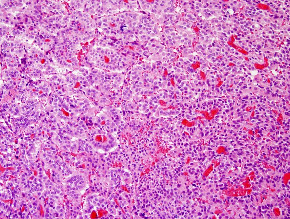
Washington University Experience | NEOPLASMS (NON-GLIAL NON-NEURONAL) | Choroid Plexus Carcinoma (CPC) | 2B2 Choroid Plexus Carcinoma (Case 2) H&E 2
2B2-4 This neoplasm is composed of complex branching fibrovascular cores lined by single to multiple layers of crowded epithelial cells, consistent with a typical appearance of choroid plexus neoplasm. The tumor however transitions into more solid to sheeted architecture with very high cellular density comprised by markedly pleomorphic cells, many of which are “monstrous” with frequent multinucleation. Mitoses (including atypical forms) are easily seen in these cellular poorly- to de-differentiated appearing foci, reaching 19/10 HPF. Focal necrosis, infiltration into the adjacent brain parenchyma, and scattered calcifications are additional notable findings.

