Table of Contents
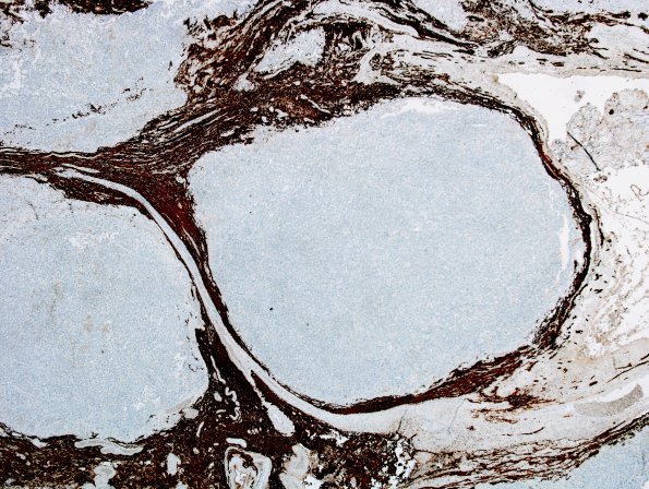
Washington University Experience | NEOPLASMS (NON-GLIAL NON-NEURONAL) | Choroid Plexus Carcinoma (CPC) | 8E Choroid Plexus Carcinoma (Case 8) GFAP.
GFAP immunohistochemistry demonstrates intense gliosis at the margins of nodules but no staining of tumor cells themselves (GFAP IHC). ---- Other immunohistochemical stains were performed (not shown). The tumor cells are focally positive for vimentin and S-100. Rare cells are positive for EMA, pancytokeratin and CAM5.2 (weak). lNl-1 Is retained in tumor and non-tumor nuclei. The tumor cells are negative for synaptophysin. P53 shows nuclear immunostaining in up to 90% of tumor cells.

