Table of Contents
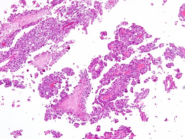
Washington University Experience | NEOPLASMS (NON-GLIAL NON-NEURONAL) | Choroid Plexus Carcinoma (CPC) | 9A1 Choroid Plexus Carcinoma (Case 9) H&E 10
Case 9 History ---- The patient is a 1 year old boy who presented with papilledema. Imaging studies revealed a large right lateral ventricle tumor. ---- 9A1-4 The tumor is a hybrid of papillary and solid-appearing areas of neoplasm with high cellularity and marked nuclear pleomorphism. Mitotic figures are readily identified, including atypical forms. Foci of micronecrosis are also seen. The tumor cells display an epithelioid cytology and are radially arranged around blood vessels. However, they are not associated with obvious fibrillary processes.

