Table of Contents
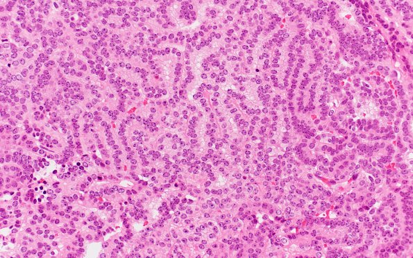
Washington University Experience | NEOPLASMS (NON-GLIAL NON-NEURONAL) | Choroid plexus papilloma | 12B1 Choroid plexus papilloma, atypical (Case 12) H&E 1
12B1-3 Sections show a neoplasm arranged in papillary pattern. These papillae are lined by a single layer of bland and uniform cuboidal to columnar epithelium with oval nuclei containing inconspicuous nucleoli, with occasional psammomatous calcifications. In multiple areas, papillae are crowded and back-to-back with focal increase in mitotic activity (2/10HPF). No solid areas, necrosis, cellular atypia, rhabdoid cells or primitive appearing cells are seen. There are areas where the tumor appears to have invaginated/invaded into gliotic brain parenchyma; however these areas may represent the base of the tumor.

