Table of Contents
Washington University Experience | NEOPLASMS (NON-GLIAL NON-NEURONAL) | Choroid plexus papilloma | 15A1 Choroid Plexus Papilloma, Atypical (Case 15) 1.jpg
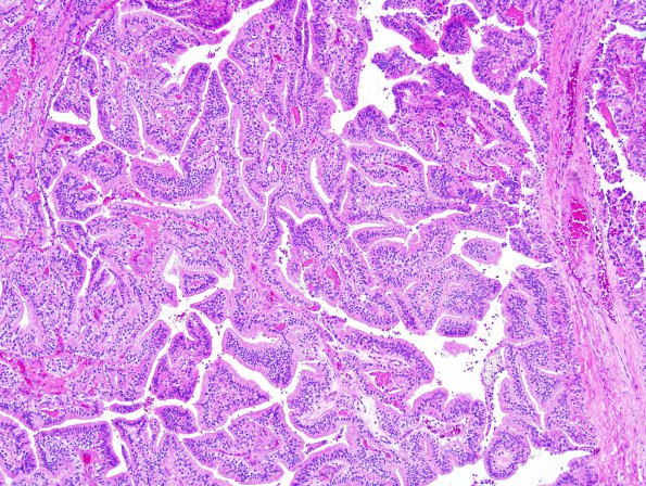
15A1-3 Sections of brain tumor show a solid, papillary neoplasm composed of delicate fibrovascular connective tissue fronds covered by a single layer of uniform columnar epithelial cells with round to oval basally situated monomorphic nuclei. Mitotic figures are identified, up to 2/10 high power fields. There is focal clear cell change. Microcalcifications are identified. No necrosis is seen.

