Table of Contents
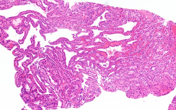
Washington University Experience | NEOPLASMS (NON-GLIAL NON-NEURONAL) | Choroid plexus papilloma | 18B1 CPP (Case 18) H&E 10X
18B1,2 Sections of the right hippocampal and lateral temporal horn ventricular lesion show a neoplasm consisting of papillary projections of tightly spaced, single layered cuboidal to columnar epithelium with vascular cores. Mitotic figures are not appreciated, and necrosis is not observed. The neoplastic cells retain a single layered monotonous appearance, and sheeting is not present.

