Table of Contents
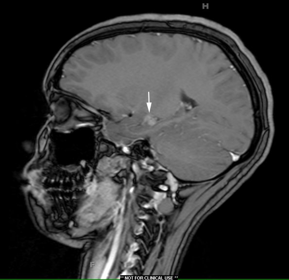
Washington University Experience | NEOPLASMS (NON-GLIAL NON-NEURONAL) | Choroid plexus papilloma | 2A1 Choroid Plexus Papilloma (Case 2) T1 W 2 copy - Copy
Case 2 History ---- The patient is a 15-year-old girl who presents with an incidentally found left temporal lobe lesion discovered in July 2014 after a fall. Initial CT shows some calcification at the side of the brain and MRI showed enhancement. She reports she continues to experience frequent (3-4 per week) headaches with photophobia and phonophobia MRI was performed revealing a T1-weighted enhancing mass concerning for ganglioglioma versus DNET versus hematoma. She denies any concerns of seizures. She denies any balance or coordination concerns. Operative procedure: Left temporal craniotomy for biopsy and lesion resection. ---- 2A1,2 A small intraventricular MRI T1-weighted contrast enhancing mass is noted in the left temporal horn in sagittal section (arrow, 2A1) and in coronal section (2A2).

