Table of Contents
Washington University Experience | NEOPLASMS (NON-GLIAL NON-NEURONAL) | Choroid plexus papilloma | 4B1 Choroid Plexus Papilloma (Case 4) H&E 2.jpg
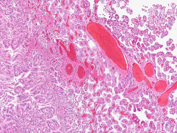
4B1,2 Histologic evaluation of the third ventricle tumor shows delicate, complex, fibrovascular connective tissue lined fronds covered by mostly a single layer of uniform cuboidal to columnar epithelioid cells. The cells are cuboidal to columnar epithelial cells with round or oval, basally situated monomorphic nuclei. In places the papillary architecture has a more compact structure. Areas at the periphery (arrowheads, B1) of the neoplasm represent collections of papillae with intercellular spaces which represent adjacent normal/non-neoplastic choroid plexus (4B2).

