Table of Contents
Washington University Experience | NEOPLASMS (NON-GLIAL NON-NEURONAL) | Choroid plexus papilloma | 6G1 CPP & multiple cysts (Case 6) cyst lining H&E 1.jpg
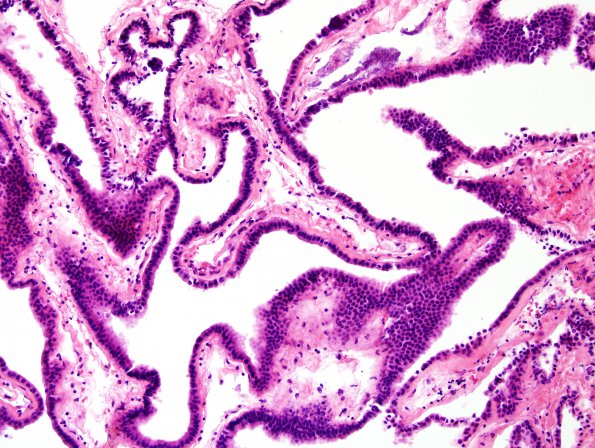
6G1-6 These images represent the histopathology of a separately biopsied cerebellar cyst lining which has collapsed as fluid leaked out. ---- 6G1 H&E stained sections of the "right cerebellar cyst" material show fragments of a single layered columnar epithelium overlying thin cores of fibrous tissue. It is difficult to tell whether true cilia are present on the epithelial surface. There are no goblet cells identified. Scattered dystrophic calcifications are present. (H&E)

