Table of Contents
Washington University Experience | NEOPLASMS (NON-GLIAL NON-NEURONAL) | Choroid plexus papilloma | 9A1 Choroid Plexus Papilloma (Case 9) -H&E_6.jpg
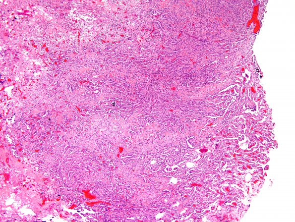
Case 9 History ---- The patient was a 20-year-old male who was diagnosed with a posterior fossa mass during an evaluation for head trauma produced by being struck by a golf club in a melee (he was a bystander). He was noted to have an occipital lesion which was resected two weeks prior. However, a CT scan showed a mass in the fourth ventricle confirmed by MRI as a homogenously enhancing mass in the fourth ventricle without obstructive hydrocephalus. He was neurologically intact with his incidentally discovered tumor. Due to its location in the fourth ventricle and the risk of obstructive hydrocephalus, the tumor was excised. Operative procedure: craniotomy and resection. ---- 9A1,2 The neoplasm is composed of multiple fragments of a papillary neoplasm composed of fibrovascular connective tissue fronds covered by a single layer of cuboidal to columnar epithelial cells with round to oval, basally situated monomorphic nuclei. Mitoses are rare. No atypical features including; increased cellularity, nuclear pleomorphism, solid growth, or areas of necrosis are seen. (H&E)

