Table of Contents
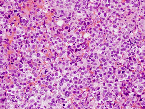
Washington University Experience | NEOPLASMS (NON-GLIAL NON-NEURONAL) | Germ Cell Neoplasm - Germinoma | 13A1 Germinoma (Case 13) H&E 3
Case 13 History ---- The patient was a ten-year-old girl with a T1 isointense, enhancing, 1.3 x 1.2 x 2 cm sellar and suprasellar mass, without radiological evidence of spinal involvement. Clinical diagnosis: Pituitary adenoma versus germinoma. Operative procedure: Transsphenoidal biopsy.
13A1,2 The neoplasm was composed of sheets of mildly discohesive, round-to-oval cells with pale, eosinophilic-to-clear cytoplasm, round-to-oval centrally-located nuclei, heterogeneously speckled chromatin, and prominent nucleoli. In an augenblick at low magnification it could be mistaken for a pituitary adenoma. However, mitotic figures are abundant. Rare small multinucleated cells with eosinophilic cytoplasm, suggestive of syncytiotrophoblasts, are also observed. However, there is no definite evidence of choriocarcinoma or other non-germinomatous germ cell tumor elements. Distributed somewhat randomly among the tumor cells is a network of fibrovascular stroma, often infiltrated by benign-appearing lymphocytes.

