Table of Contents
Washington University Experience | NEOPLASMS (NON-GLIAL NON-NEURONAL) | Germ Cell Neoplasm - Germinoma | 9A Germinoma & Yolk Sac (Case 9) 1 copy
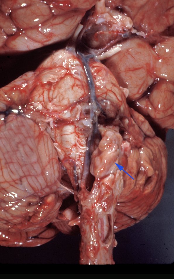
Case 9 History ---- This patient was a 16 year old female with a history of resection of a suprasellar “ectopic pinealoma”, an archaic term for germinoma, in 1967. In 1968 a metastatic pinealoma was resected from the thoracic spinal cord. In 1969 a Holter valve was place for hydrocephalus. Her condition continued to worsen and she died in August 1970. ---- At autopsy the unfixed brain weighed 1400g. The leptomeninges overlying the cerebral hemispheres appear slightly thickened over the convexities and at the base of the brain with visible nodules of tumor (arrow) along the brainstem and optic chiasm. Coronal sections of the cerebral hemispheres showed a normal appearing white and gray matter. Examination of the spinal cord revealed extradural tumor at the lumbar levels of the spinal cord which extended into the adjacent vertebral column. ---- 9A This view of the ventral aspect of the brain shows a large extrapontine mass (arrow) and the optic chiasm as well as a gelatinous appearance of the leptomeninges over the temporal lobe and cerebellum.

