Table of Contents
Washington University Experience | NEOPLASMS (PINEAL) | PPTID (Pineal Parenchymal Tumor Intermediate Differentiation) | 2A3 PPTID (Case 2) H&E 8.jpg
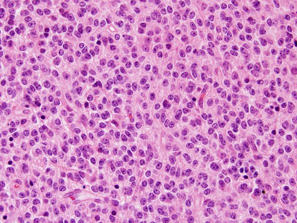
Tumor cell density is high in this tumor that is primarily solid but which focally appears to invade white matter. There is no evidence of necrosis or microvascular proliferation. Calcification is noted focally. Tumor cells contain uniform round nuclei with small nucleoli; associated cytoplasm is ill-defined and generally eosinophilic. A subset of tumor nuclei are surrounded by clear halos. Mitotic figures are generally scattered, however, occasionally two or three are seen in a single high power field. Focally, both Homer Wright rosettes, pineocytomatous and Flexner Wintersteiner rosettes are seen. There is no evidence of degenerative atypia or ganglionic differentiation. (H&E)

