Table of Contents
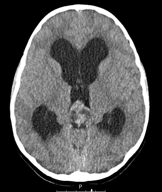
Washington University Experience | NEOPLASMS (PINEAL) | Papillary Tumor Pineal Region (PTPR) | 1A1 Papillary Tumor Pineal Region (PTPR, Case 1) CT A - Copy
Case 1 History ---- The patient is a 7-year-old girl who presented after several months of headache, visual changes, and two generalized tonic-clonic seizures. Computed tomography at an OSH revealed a pineal region tumor approximately 2.6 cm in largest dimension causing obstructive hydrocephalus and triventricular ventriculomegaly. MRI at BJH showed a heterogeneously enhancing pineal lesion. Radiological impression: Germ cell tumor versus primary pineal neoplasm. Operative procedure: Endoscopic third ventriculostomy with biopsy. ---- 1A1 A preoperative CT scan shows a pineal mass and obstructive hydrocephalus.

