Table of Contents
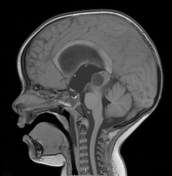
Washington University Experience | NEOPLASMS (PINEAL) | Papillary Tumor Pineal Region (PTPR) | 2A1 Papillary Tumor of the Pineal Region (Case 2) T1noC sag - Copy
Case 2 History ---- The patient is a 22-month-old girl with a history of weeks of gait difficulty and incoordination. MRI shows marked obstructive hydrocephalus and a 1.8 x 1.8 x 2.3 cm partially solid, partially cystic, enhancing mass without calcifications, centered in the pineal region, near the posterior aspect of the third ventricle. The largest cyst, located anteriorly, measures 1.5 cm. Radiographic impression is an origin from the pineal gland as opposed to the tectum. No enhancing spinal lesions are identified. Operative procedure: Endoscopic third ventriculostomy and biopsy of tumor. ---- 2A1,2 This T1-weighted sagittal scan demonstrates a partly cystic pineal mass which enhances with contrast.

