Table of Contents
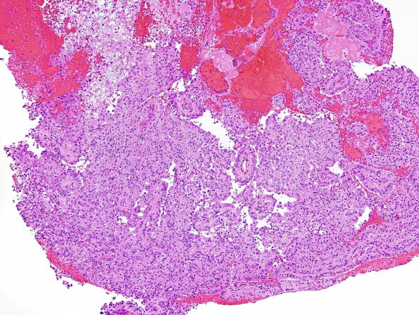
Washington University Experience | NEOPLASMS (PINEAL) | Papillary Tumor Pineal Region (PTPR) | 2B1 Papillary Tumor of the Pineal Region (Case 2) H&E 12.jpg
2B1-4 The specimen consists of multiple fragments of a cellular neoplasm associated with a thick fibrous capsule consistent with cyst wall. The tumor cells are generally epithelioid, with a moderate amount of pale eosinophilic cytoplasm, thin crisp eosinophilic borders, bland oval nuclei, fine chromatin and inconspicuous nucleoli. These cells appear in irregularly paved sheets and, more distinctively, in short, poorly-formed columnar epithelia, focally around large papillary-like structures. Mitotic figures are not identified. Pink amorphous material intermixed with blood is present in several foci, consistent with degenerative cyst contents/necrotic debris.

