Table of Contents
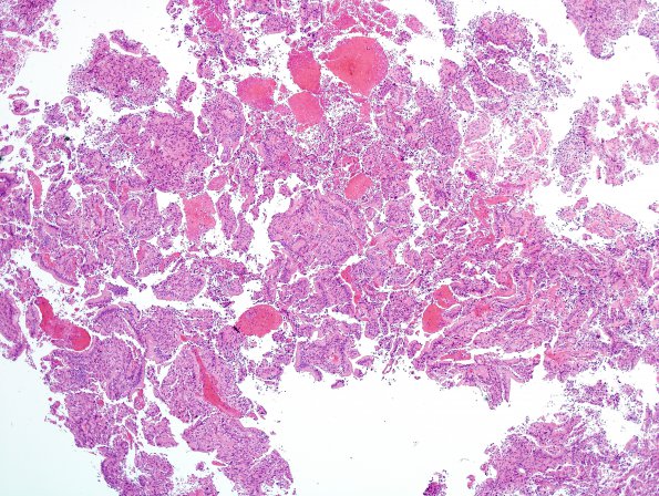
Washington University Experience | NEOPLASMS (PINEAL) | Papillary Tumor Pineal Region (PTPR) | 5A1 Papillary Tumor Pineal Region (Case 5) H&E 11.jpg
Case 5 History ---- The patient is a 37 year old man who has had headaches and visual loss for two years. Imaging studies reveal a pineal region mass with associated obstructive hydrocephalus. ---- 5A1-6 Sections reveal an epithelioid neoplasm arranged in both papillary and solid configurations. The tumor has a relatively sharp demarcation with adjacent fragments of non-neoplastic pineal parenchyma. There is mild to moderate nuclear pleomorphism, with the majority of tumor cells appear as large columnar epithelioid cells arranged in a papillary configuration and focally forming gland-like or rosette-like structures with central lumens. The tumor cells have oval nuclei with small nucleoli and abundant clear to lightly eosinophilic cytoplasm. Mitotic figures are rare but present. There was no definite evidence of tumor necrosis. (H&E)

