Table of Contents
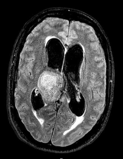
Washington University Experience | NEOPLASMS (PINEAL) | Papillary Tumor Pineal Region (PTPR) | 7A1 Papillary Tumor Pineal Region (Case 7) 3D FLAIR axial - Copy
Case 7 History ---- The patient is a 34-year-old man with a midbrain/pineal region mass. Stereotactic biopsy was performed at an outside hospital in mid-January 2014 and reviewed here in consultation, yielding the diagnosis 'papillary tumor of the pineal region. MRI 2 months later shows a ~6.5 x 4.6 x 5 cm T1 and T2 heterogeneous, mildly enhancing mass with irregular internal cystic spaces, arising from the region of the pineal gland. The tumor extends superiorly, exerting mass effect on the third ventricle and right lateral ventricle, and also extends anteriorly, into the suprasellar cistern. Operative procedure: Craniotomy for resection with intraoperative MRI. 7A1-4 Axial MRI Studies: 7A1 The tumor is hyperintense on this FLAIR scan.

