Table of Contents
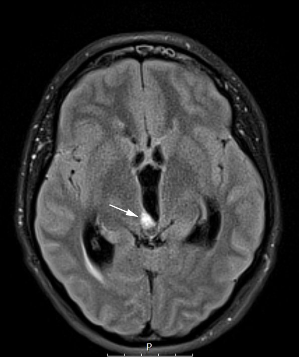
Washington University Experience | NEOPLASMS (PINEAL) | Papillary Tumor Pineal Region (PTPR) | 9A1 Papillary Tumor Pineal Region (Case 9) FLAIR axial A copy - Copy
Case 9 History ---- The patient is a 28 year-old woman who presented with progressive headaches and visual difficulties. MRI of the brain showed a 1.1 x 0.8 x 1.0 cm rounded heterogenous lesion along the posterior right lateral wall of the distal third ventricle, anterior and inferior to the expected location of the pineal gland. The lesion displaces the tectum posteriorly and obstructs the cerebral aqueduct. The patient underwent a ventriculostomy that was aborted due to a small amount of bleeding obstructing the field of view for biopsy. Operative procedure: Reexploration with endoscopic biopsy of third ventricular lesion. ---- MRI Studies: 9A1 The lesion is quite small (arrow) in this FLAIR scan.

