Table of Contents
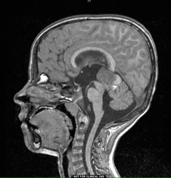
Washington University Experience | NEOPLASMS (PINEAL) | Pineoblastoma | 2A2 Pineoblastoma (Case 2) T1 no contrast - Copy
2A2 This T1-weighted scan shows a well-defined, multi-lobulated, uniformly-enhancing 3.4-cm mass, with internal hemorrhage and calcification, centered in the pineal region, with extension anteriorly into the third ventricle and posteriorly into the supra-vermian cistern.

