Table of Contents
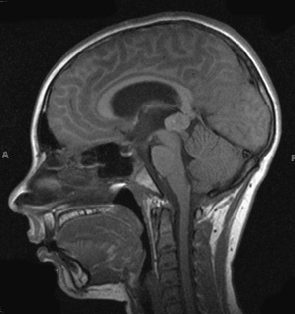
Washington University Experience | NEOPLASMS (PINEAL) | Pineoblastoma | 4A1 Pineoblastoma (Case 4) T1 3 - Copy
Case 4 History ---- The patient is an 11-year-old boy who initially presented with headache, nausea, emesis, and diplopia. MRI shows a solid and cystic, heterogeneously post-contrast enhancing mass with calcifications located in the pineal region. Operative procedure: Craniotomy and excision. 4A1-6 MRI studies: 4A1,2 A T1-weighted scan without (4A1) and with (4A2) contrast enhancement.

