Table of Contents
Washington University Experience | NEOPLASMS (PINEAL) | Pineocytoma | 10A3 Pineocytoma (Case 10) H&E 5.jpg
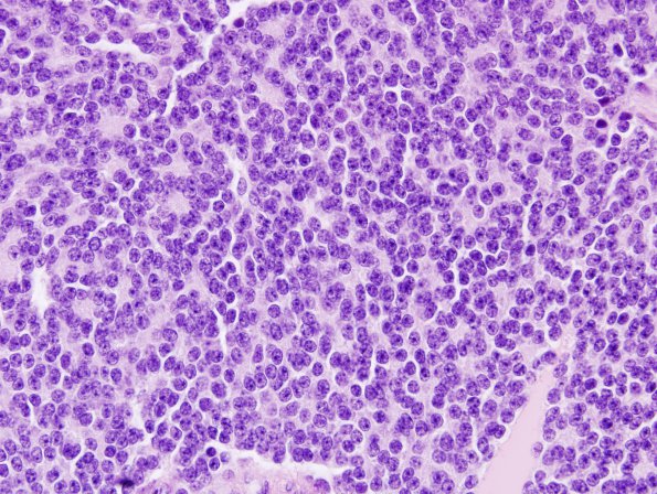
Tumor cell density is high in this well vascularized neoplasm. Small neurocytic rosettes are scattered throughout the tumor, which itself is populated by small round monotonous nuclei bearing ill-defined eosinophilic cytoplasm. Only occasional mitotic figures are identified. Although foci of calcification are noted, native pineal parenchymal tissue cannot be discerned. ---- Ancillary data (not shown): Synaptophysin is strongly positive, and there is focal neurofilament protein positivity. The Ki-67 proliferative index is generally low.

