Table of Contents
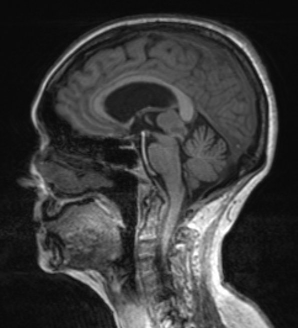
Washington University Experience | NEOPLASMS (PINEAL) | Pineocytoma | 1A1 Pineocytoma (Case 1) T1 MPRAGE 2 - Copy
Case 1 History ---- The patient is a 72-year-old woman who noted impaired balance and ability to walk approximately nine months previously which continued to worsen. MR imaging showed a 2.5 x 1.9 x 2.0 cm mass centered near the opening of the cerebral aqueduct within the third ventricle. The mass showed slight T1 hypointensity, T2 isointensity, and enhancement. The pineal gland was not definitively seen distinct from the mass. Operative procedure: Endoscopic third ventriculostomy. ---- 1A1,2 T1-weighted sagittal section without (1A1) and with contrast (1A2)

