Table of Contents
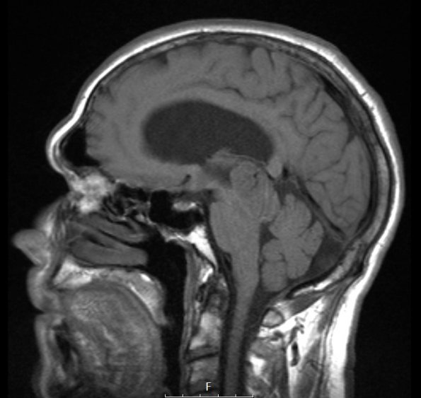
Washington University Experience | NEOPLASMS (PINEAL) | Pineocytoma | 5A1 Pineocytoma (Case 5) T1 IPAT noC - Copy
Case 5 History ---- The patient is a 66 year old man with a pineal region tumor, incidentally identified in January 2004 during evaluation of right facial and dental pain. A ventricular shunt was placed in August 2005 to relieve obstructive hydrocephalus caused by the tumor. A few months prior to admission, he experienced occasional bifrontal headaches, diplopia, rare dizziness, and gait abnormality. MRI performed in October 2006 shows a 3.7cm enhancing, hemorrhagic mass in the pineal region that has demonstrated little change in size. Operative procedure: Endoscopic biopsy of pineal region tumor. ---- 5A1,2 This T1-weighted sagittal scan shows an isointense mass (5A1) which enhances with contrast (5A2).

