Table of Contents
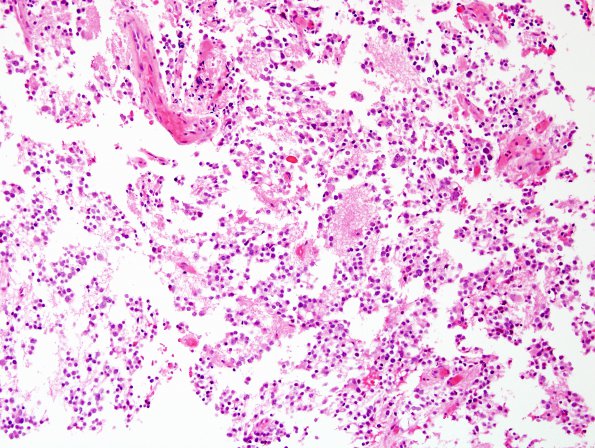
Washington University Experience | NEOPLASMS (PINEAL) | Pineocytoma | 7A1 Pineocytoma (Case 7) H&E 2.jpg
Case 7 History ---- The patient is a 52 year old woman with a brightly enhancing pineal region mass, which appeared stable on serial MRI studies. ---- 7A1,2 Sections reveal a moderately cellular, relatively solid-appearing neoplasm composed predominantly of small to medium size nuclei with bland chromatin, salt and pepper chromatin, and either clear perinuclear halos or small quantities of eosinophilic cytoplasm. Occasional cells are enlarged and contain bizarre nuclei, most consistent with degenerative atypia given the lack of associated mitotic activity. In some areas, the tumor cells are arranged around central aggregates of delicate fibrillary material, resembling neuropil. These structures are consistent with pineocytic rosettes. Mitotic figures are hard to find and there is no evidence of microvascular proliferation or necrosis.

