Table of Contents
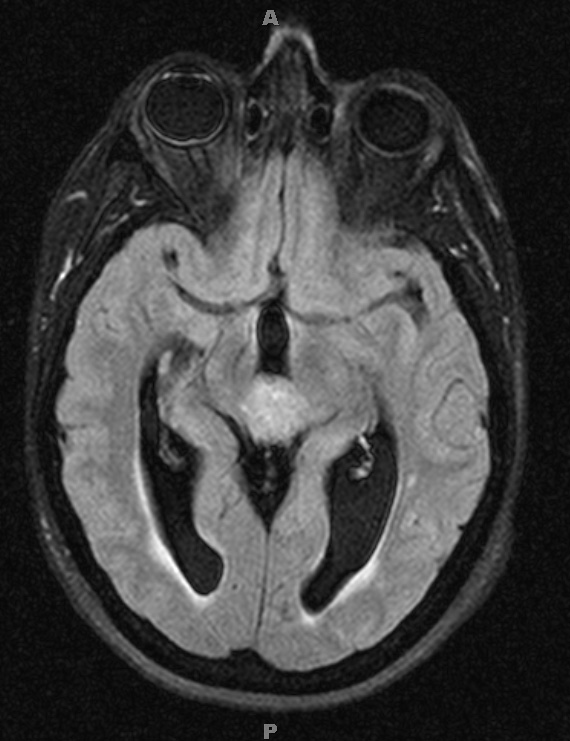
Washington University Experience | NEOPLASMS (PINEAL) | Pineocytoma | 8A1 Pineocytoma (Case 8) FLAIR - Copy
Case 8 History ---- The patient is a 51-year-old woman who developed lightheadedness. MRI showed a 2.5 cm pineal region mass that obstructed the cerebral aqueduct causing hydrocephalus. The radiographic differential diagnosis included pineocytoma, pineal parenchymal tumor of indeterminate differentiation, papillary tumor of the pineal region and less likely germinoma or pineoblastoma. Operative procedure: Endoscopic third ventriculostomy and biopsy of pineal lesion. ---- 8A1-5 MRI studies: 8A1 The tumor is hyperintense compared to adjacent brain in this FLAIR scan.

