Table of Contents
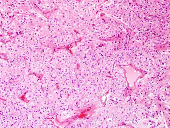
Washington University Experience | NEOPLASMS (PINEAL) | Pineocytoma | 9A2 Pineocytoma w neuronal diffn (Case 9) H&E 1.jpg
Microscopic sections reveal a pineocytoma with extensive neuronal differentiation. This tumor has a lobular low power architecture that forms a discrete border with adjacent gliotic brain. Tumor cells at times cluster, and are embedded within a rich neuropil-like background. Pineocytomatous rosettes are identified. There is a range of neuronal differentiation which includes neurocytes, large ganglion cells, and intermediate sized ganglioid cells. Numerous multinucleate ganglion cells are seen, some of which are quite large and vacuolated. There is no necrosis or microvascular proliferation, and mitoses are rare. Degenerative type changes include enlarged naked hyperchromatic nuclei, several hyalinized blood vessels, and scattered foci of hemosiderin deposition.

