Table of Contents
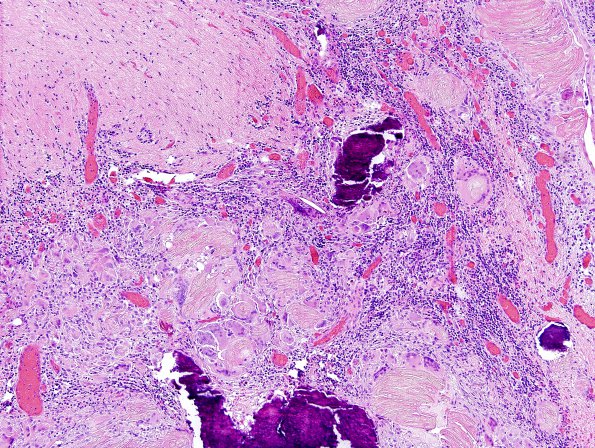
Washington University Experience | NEOPLASM (SELLAR) | Craniopharyngioma, adamantinomatous | 14A1 Cranio SP resection, no live tumor (Case 14) H&E 3.jpg
14A1-4 Sections show fragments of glial tissue involved by a predominantly chronic inflammatory infiltrate, composed of plasma cells and lymphocytes, a minor component of acute inflammatory cells composed of neutrophils and eosinophils, multinucleated giant cells, and most notably, fragments of wet keratin as well as large, coarse calcifications. The adjacent glial tissues show numerous Rosenthal fibers, consistent with "piloid gliosis." There is no evidence of either a stratified squamoid epithelium or stellate reticulum to suggest viable tumor is present in these sections. Some of the multinucleated giant cells contain fragments of engulfed wet keratin, representing a chronic process and suggestive of residual non-neoplastic elements or ruptured cyst contents consistent with the patient's previously documented craniopharyngioma.

