Table of Contents
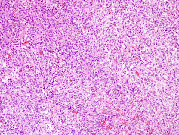
Washington University Experience | NEOPLASMS - CRANIAL AND PARASPINAL NERVEs | Malignant Peripheral Nerve Sheath Tumor (MPNST) | 11E1 MPNST (Case 11) recurrence H&E 4
Case 11 recurrence 3+ years later. ---- Following his previous resection of the tumor located near the lateral wall of the right cavernous sinus he underwent gamma knife radiotherapy in 8/2009, and subsequently received adjuvant chemotherapy with ifosfamide and epirubicin. MRI studies in April and August of 2012 showed a new dural-based 1.8 x 1 x 1.5 cm enhancing mass in the left prepontine cistern (which was also targeted with gamma knife radiotherapy in May 2012) and an additional enhancing 1.4 x 1 x 1.1 cm mass with fluid/fluid levels and internal hemosiderin, located in the midbrain (superior colliculus), abutting the cerebellar vermis. In 09/2012 and 10/2012, he presented with symptoms associated with hydrocephalus and acute hemorrhage within the midbrain/cerebellar lesion. Operative procedure: Posterior fossa craniotomy and tumor excision. ---- 11E1,2 Sections of the "posterior fossa mass" showed multiple fragments of neoplastic tissue formed predominantly by sheets of epithelioid tumor cells with moderately pleomorphic oval nuclei, prominent nucleoli, and variably eosinophilic or coarsely vacuolated/cleared cytoplasm. Some lesser areas show spindled morphology. Other focal areas show increased cellularity, finely speckled dense chromatin, small pseudorosettes and papillary architecture. Mitotic figures are common. Focal areas of necrosis are observed. The interface with brain parenchyma appears broad rather than diffusely infiltrative, and shows robust reactive changes as well as abundant hemosiderin deposits.

