Table of Contents
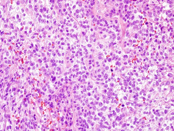
Washington University Experience | NEOPLASMS - CRANIAL AND PARASPINAL NERVEs | Malignant Peripheral Nerve Sheath Tumor (MPNST) | 11E2 MPNST (Case 11) recurrence H&E 1
Sections showed multiple fragments of neoplastic tissue formed predominantly by sheets of epithelioid tumor cells with moderately pleomorphic oval nuclei, prominent nucleoli, and variably eosinophilic or coarsely vacuolated/cleared cytoplasm. Some lesser areas show spindled morphology. Other focal areas show increased cellularity, finely speckled dense chromatin, small pseudorosettes and papillary architecture. Mitotic figures are common. Focal areas of necrosis are observed. The interface with brain parenchyma appears broad rather than diffusely infiltrative, and shows robust reactive changes as well as abundant hemosiderin deposits.

