Table of Contents
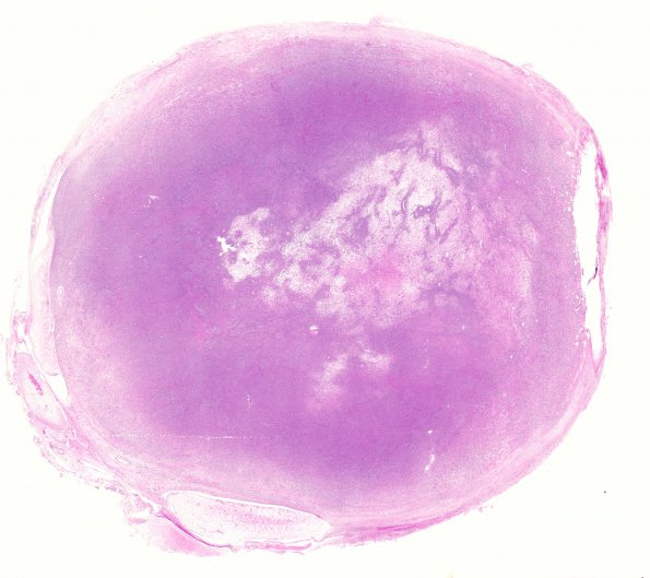
Washington University Experience | NEOPLASMS - CRANIAL AND PARASPINAL NERVEs | Malignant Peripheral Nerve Sheath Tumor (MPNST) | 15A1 MPNST (Case 15) H&E WM
Case 15 History ---- The patient is a 31-year-old man with a clinical history of neurofibromatosis, who has found to have a palpable mass in the right femoral region. Ultrasound showed a 4.6 x 3.0 x 2.7 cm hypoechoic mass. The patient underwent excision of the right femoral/inguinal lesion (this specimen).
15A1-4 Hematoxylin and eosin stained sections of the right femoral/inguinal lesion show a high-grade, well-circumscribed, hypercellular spindle cell neoplasm. The tumor cells have atypical ovoid nuclei with marked pleomorphism, coarse chromatin, and occasional prominent nucleoli. The tumor has a herringbone growth pattern in areas, and occasional paucicellular regions with an eosinophilic proteinaceous background. Areas reminiscent of classic neurofibroma, with interspersed thick and thin collagen fibers, are seen. Mitotic activity is elevated, with 26 mitoses/10 HPF. Multifocal areas of necrosis are present comprising ~5% of the tumor volume.

