Table of Contents
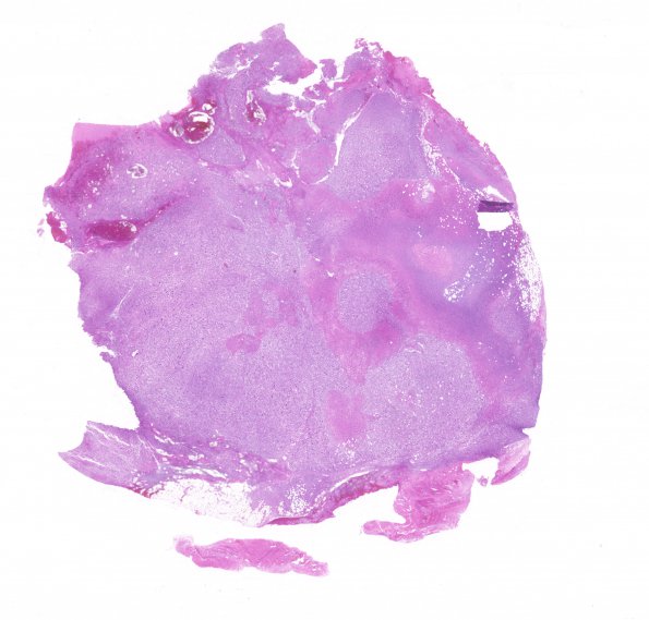
Washington University Experience | NEOPLASMS - CRANIAL AND PARASPINAL NERVEs | Malignant Peripheral Nerve Sheath Tumor (MPNST) | 18B1 MPNST, plex NF in NF1, epitheloid (Case 18) H&E WM
Two histological patterns of neoplasia are seen. ---- One pattern, representing neurofibroma and present within all the specimens included in this case, is generally modestly cellular; its wavy spindled cells with oval to elongated nuclei show only very rare mitotic activity and appear within a variably edematous/myxoid background of wavy collagen fibrils. ---- In a few areas, the neurofibroma tissue exhibits increased cellularity. The second histological pattern is formed by a markedly pleomorphic and highly proliferative cell population with many areas of necrosis. The cells range from smaller spindled forms to large, irregular epithelioid cells with one or more strikingly pleomorphic nuclei, coarse chromatin, occasional nuclear pseudoinclusions, prominent nucleoli, and variably eosinophilic or amphophilic cytoplasm. Sections from the sacral mass specimen show evidence of bone and soft tissue invasion by the malignant tumor tissue, accompanied by lytic and remodeling changes in the bone.

