Table of Contents
Washington University Experience | NEOPLASMS - CRANIAL AND PARASPINAL NERVEs | Malignant Peripheral Nerve Sheath Tumor (MPNST) | 20A1 Neurofibromatosis 1 (Case 20) 1 copy
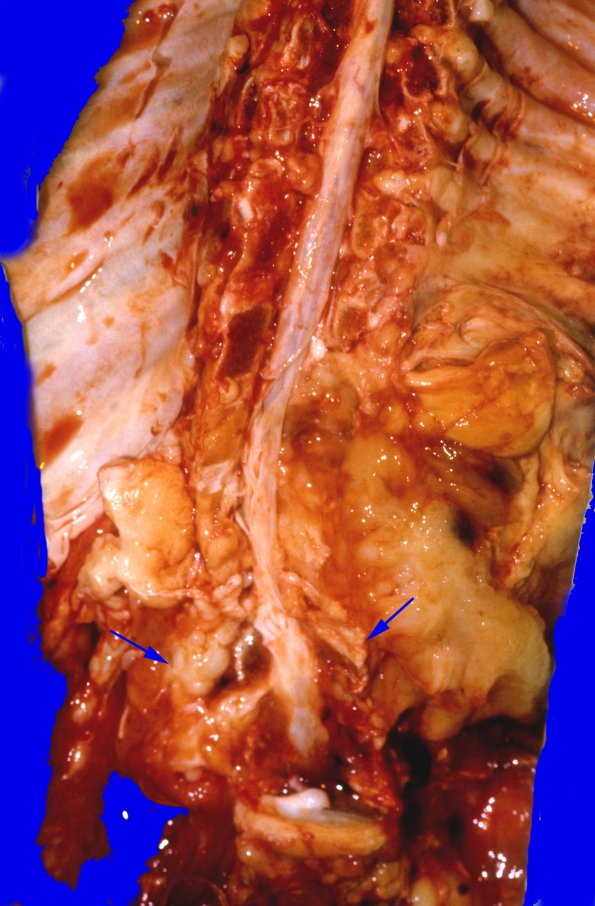
Examination of the external and cut surfaces of the spinal cord, as well as the spinal dura discloses extensive enlargement of the spinal nerve roots as they emerge from the dura (arrows). At some points, these roots measure up to 2 cm. in diameter. The enlargement is associated with effacement of the normal anatomical markings on the cut surfaces of the nerve roots and only a few fascicles can be identified grossly. For the most part, the normal fascicles have been replaced by firm, rubbery, white, or greyish-white tissue. Similar changes are seen in nerve trunks throughout the body.

