Table of Contents
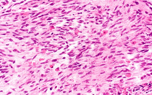
Washington University Experience | NEOPLASMS - CRANIAL AND PARASPINAL NERVEs | Malignant Peripheral Nerve Sheath Tumor (MPNST) | 4A2 MPNST (Case 4) H&E 3
Hematoxylin and eosin stained sections of the resection show a tumor composed of alternating hypercellular fascicles and less cellular fibrous to myxoid regions. The hypercellular areas consist of crowded, variably atypical spindled cells with hyperchromatic, tapered nuclei, numerous mitoses (up to 10/10 HPF) and apoptotic bodies. Focal areas of geographic necrosis are present. There are foci of tumor cells with epithelioid differentiation, arranged in small whorled nests. Rare individual rhabdomyoblastic cells are present, as are areas of metaplastic osseous differentiation. The less cellular areas demonstrate minimally atypical, elongated, tapered/pointed, wavy, or buckled nuclei characteristic of lower grade nerve sheath tumors.

