Table of Contents
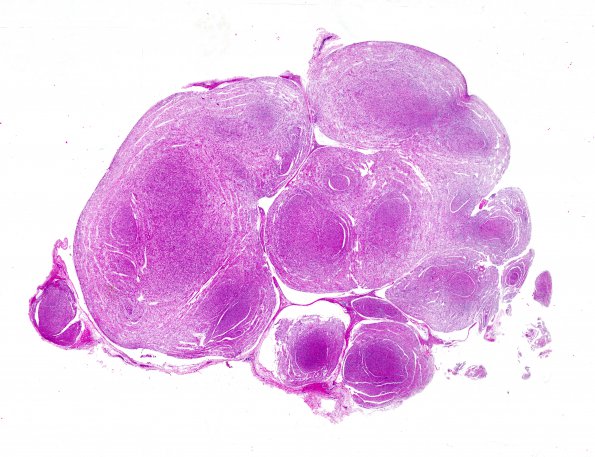
Washington University Experience | NEOPLASMS - CRANIAL AND PARASPINAL NERVEs | Neurofibroma | 14A1 Neurofibroma, plexiform (Case 14) H&E whole mount 2
Case 14 History ---- The patient is a 26 year old man who was diagnosed with neurofibromatosis type 1 at the age of 12 when a large tumor was removed from his neck. He did not have difficulty for the next 14 years but subsequently developed a large tumor in his abdomen that wrapped around his aorta. Additionally, repeat CT of his neck showed the original tumor had recurred. ---- 14A1-4 The provided sections show a neoplasm that involves and expands multiple associated fascicles of peripheral nerve. The neoplasm, well circumscribed by perineurium, is composed of elongated small spindled cells with hyperchromatic, thin, tapered nuclei and interspersed bundles of collagen, embedded within a mucinous matrix. Cell density ranges from low to moderate. There is no evidence of significant mitotic density, or necrosis. (H&E)

