Table of Contents
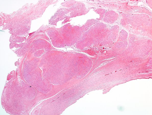
Washington University Experience | NEOPLASMS - CRANIAL AND PARASPINAL NERVEs | Neurofibroma | 15 Neurofibroma, plexiform, hyalinized (Case 15) H&E 1
Case 15 History ---- The patient is a 43 year old man with a well -circumscribed, homogenously enhancing mass in the right L4-L5 neural foramen that is radiologically indistinguishable from the existing L4 nerve root. ---- 15A Sections of the L4 foramen mass show a fascicular arrangement of hyalinized tissue with a hint of a perineurial surround of each fascicle.
Not shown: The immunohistochemical stains performed show reactive vimentin with a nuclear pattern, and non-reactive neurofilament and EMA. Interpretation of S100 is equivocal due to histotechnical difficulties. A Bielschowsky stain shows scattered darkly stained material which are not compelling axons. There is no evidence of Schwannoma or metastatic carcinoma. These features are more consistent with a plexiform neurofibroma with extensive hyalinization.

