Table of Contents
Washington University Experience | NEOPLASMS - CRANIAL AND PARASPINAL NERVEs | Neurofibroma | 18B1 Neurofibroma, plexiform (Case 18)
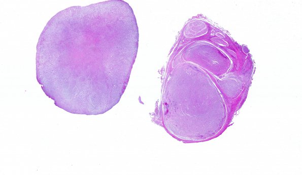
18B1-4 Histological sections of the "foraminal mass" show a radially well-circumscribed neoplasm involving multiple fascicles with an intrafascicular growth pattern. The tumor is composed of a mixed population of cells with mildly pleomorphic nuclei, ranging from spindled to round/oval, intermixed with modestly thick collagen bundles; all of these are randomly arranged within a pale myxoid background. Occasional larger, irregular nuclei are present, consistent with those of Schwann cells, but are not suggestive of atypia. Occasional small clusters of benign-appearing lymphocytes are also identified. Subtle but abundant, regularly-spaced, slit-like vessels are evident. Rare foci of tumor tissue appear hyalinized and hypocellular. Ganglion cells with large round nuclei, prominent nucleoli and abundant speckled amphophilic cytoplasm occasionally appear within the tumor parenchyma, but are more prominent in the periphery of the tumor, adjacent to the fibrous pseudocapsule; this pattern provides evidence that the tumor has an expansile infiltrating growth pattern. Normal-appearing nerve fascicles with little or no involvement by tumor are also observed within the fibrous pseudocapsule. Mitotic figures appear at a density that focally ranges up to 3/10HPFs, but that is almost uniformly less than 1/10HPF. No areas of hypercellularity, cellular crowding, uniformly enlarged nuclei, necrosis, or aberrant differentiation are present to suggest malignant transformation. No Antoni A / Antoni B tissue patterns or Verocay bodies are present to suggest a diagnosis of schwannoma.

