Table of Contents
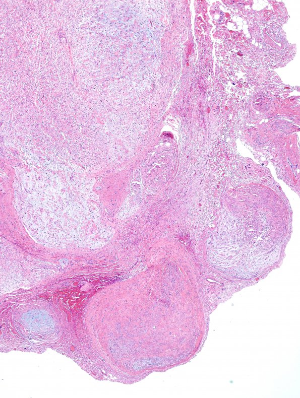
Washington University Experience | NEOPLASMS - CRANIAL AND PARASPINAL NERVEs | Neurofibroma | 6C2 Neurofibroma, plexiform (NF1) (Case 6) H&E 3
6C1-6 The neoplastic cells are small, with hyperchromatic elongated nuclei, and are often associated with dense bundles of collagen fibers. These collagen bundles are somewhat haphazardly dispersed within pale myxoid material, in contrast with the nerves' eosinophilic axonal processes, which are uniformly parallel and show greater preservation within the central regions of the fascicles. The tumor cell cytoplasm is minimal and eosinophilic; cell processes are thin and are often imperceptible. Vessels associated with the neoplastic tissue do not show hyalinization. Significant mitotic activity is not appreciated. There is no evidence of hypercellularity or necrosis. This histological pattern, present in each specimen, describes neurofibroma; the presence of each of these tumors in multiple associated fascicles defines them as plexiform neurofibromas. Features concerning for malignant transformation are not identified.

