Table of Contents
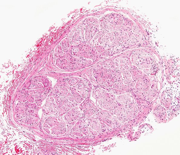
Washington University Experience | NEOPLASMS - CRANIAL AND PARASPINAL NERVEs | Perineurioma, Intraneural | 1A1 Perineurioma (Case 1) H&E 1.jpg
1A1-3 Microscopic examination of routinely stained sections of paraffin-embedded left peroneal nerve biopsy material showed cross sections of abnormal peripheral nerve. The perineurial fibrous tissue appears thickened, and surrounds fascicles which are themselves divided into mini-fascicles by septae proceeding from the perineurium. These mini-fascicles contain a reduced density of large and small myelinated axons, as well as multiple concentrically-lamellated, mildly basophilic 'onion-bulb'-like structures. Most of these 'onion-bulbs' surround one or more thinly myelinated axons.

